„”
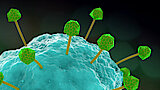
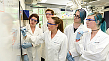
Furthermore, we characterize structure and dynamics of bacterial supramolecular assemblies. For example, we have determined the structure of the bactofilin BacA by solid-state NMR. Bactofilins are a new class of cytoskeletal proteins that are involved in key cellular processes. For instance, in the human pathogen Helicobacter pylori, they are responsible for maintaining its characteristic helical cell shape, a feature required for cells to efficiently colonize the gastric mucus. We discovered that bactofilins adopt a β-helical architecture (see Figure), which has not been observed before for other cytoskeletal filaments. Interestingly, however, the structure bears similarities to that of the fungal prion protein HET-s. (Vasa et al., PNAS 2015 and Shi et al., Science Advances 2015). We also introduced a general hybrid approach for determining the structures of supramolecular assemblies. Cryo-electron microscopy (cryo-EM) data define the overall envelope of the assembly and rigid-body orientation of the subunits while solid-state NMR chemical shifts and distance restraints define the local secondary structure, protein fold and inter-subunit interactions. Using this approach we could determine the structure of the type-III secretion system needle of Shigella flexneri to a very high precision (Demers et al., Nature Communications 2014; see also our previous work: Loquet et al., Nature 2012). Recently, our lab succeeded in solving the structure of a large and flexible supramolecular assembly: the SPP1 bacteriophage tail tube. Using tailored proton-detected 4D experiments, also developed by our lab (e.g. Zinke et al., Angewandte Chemie 2017), the complex system could be readily investigated. Distance restraints from solid-state NMR were combined with cryo-EM yielding an atomic-resolution structure. Additionally, the dynamics of the system were assessed revealing a spinal column architecture with rigid hexameric rings connected by flexible linkers (Zinke et al., Nature Communications 2020).

High-resolution 3D structure of the cytoskeletal protein BacA (Shi et al., Science Advances 2015).
The Leibniz-Forschungsinstitut für Molekulare Pharmakologie (FMP) is part of the Forschungsverbund Berlin e.V. (FVB), which legally represents seven non-university research institutes - members of the Leibniz Association - in Berlin.
Leibniz-Forschungsinstitut für Molekulare Pharmakologie im Forschungsverbund Berlin e.V. (FMP)
Campus Berlin-Buch
Robert-Roessle-Str. 10,
13125 Berlin, Germany